Showing posts with label Human Body. Show all posts

Thursday, 17 April 2014

What do the enzymes inside a cell do?

What do the enzymes inside a cell do?


Even the tiniest microscopic bacteria cell can have about 1,000 enzymes at work inside.  Enzymes are what make all the chemical reactions in the cell possible.  The human body is made up of trillions of cells, and there are different cells for different functions.  Cells are little bundles of chemical reactions. They reproduce, they create energy, and they break molecules down and build them up. All that action going on is made possible by the enzymes. 

Enzymes are proteins inside the cells. They are formed by special chains of amino acids that come together in different shapes to do special jobs, like breaking down sugar and fat molecules or to make more enzymes. The cells need the enzymes to live, and each different enzyme has its own work to do.  When the body is missing a type of enzyme, the cells can't work properly. This can lead to problems for the person whose body is missing the enzymes.

Wednesday, 2 April 2014

How does your brain stores information?


Storage Of Information In our Brain

The more you know about your memory, the better you'll understand how you can improve it. Here's a basic overview of how your memory works and how aging affects your ability to remember.
Your baby's first cry...the taste of your grandmother's molasses cookies...the scent of an ocean breeze. These are memories that make up the ongoing experience of your life -- they provide you with a sense of self. They're what make you feel comfortable with familiar people and surroundings, tie your past with your present, and provide a framework for the future. In a profound way, it is our collective set of memories -- our "memory" as a whole -- that makes us who we are.
Most people talk about memory as if it were a thing they have, like bad eyes or a good head of hair. But your memory doesn't exist in the way a part of your body exists -- it's not a "thing" you can touch. It's a concept that refers to the process of remembering.
In the past, many experts were fond of describing memory as a sort of tiny filing cabinet full of individual memory folders in which information is stored away. Others likened memory to a neural supercomputer wedged under the human scalp. But today, experts believe that memory is far more complex and elusive than that -- and that it is located not in one particular place in the brain but is instead a brain-wide process.
Do you remember what you had for breakfast this morning? If the image of a big plate of fried eggs and bacon popped into your mind, you didn't dredge it up from some out-of-the-way neural alleyway. Instead, that memory was the result of an incredibly complex constructive power -- one that each of us possesses -- that reassembled disparate memory impressions from a web-like pattern of cells scattered throughout the brain. Your "memory" is really made up of a group of systems that each play a different role in creating, storing, and recalling your memories. When the brain processes information normally, all of these different systems work together perfectly to provide cohesive thought.
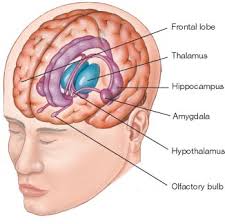
What seems to be a single memory is actually a complex construction. If you think of an object -- say, a pen -- your brain retrieves the object's name, its shape, its function, the sound when it scratches across the page. Each part of the memory of what a "pen" is comes from a different region of the brain. The entire image of "pen" is actively reconstructed by the brain from many different areas. Neurologists are only beginning to understand how the parts are reassembled into a coherent whole.
If you're riding a bike, the memory of how to operate the bike comes from one set of brain cells; the memory of how to get from here to the end of the block comes from another; the memory of biking safety rules from another; and that nervous feeling you get when a car veers dangerously close, from still another. Yet you're never aware of these separate mental experiences, nor that they're coming from all different parts of your brain, because they all work together so well. In fact, experts tell us there is no firm distinction between how you remember and how you think.
This doesn't mean that scientists have figured out exactly how the system works. They still don't fully understand exactly how you remember or what occurs during recall. The search for how the brain organizes memories and where those memories are acquired and stored has been a never-ending quest among brain researchers for decades. Still, there is enough information to make some educated guesses. The process of memory begins with encoding, then proceeds to storage and, eventually, retrieval.

Storage of Information:

The brain stores memories in two ways. Short-term memories like a possible chess move, or a hotel room number are processed in the front of the brain in a highly developed area called the pre-frontal lobe, according to McGill University and the Canadian Institute of Neurosciences, Mental Health and Addiction.
Short-term recollection is translated into long-term memory in the hippocampus, an area in the deeper brain. According to McGills , the hippocampus takes simultaneous memories from different sensory regions of the brain and connects them into a single "episode" of memory, for example, you may haveone memory of a dinner party rather than multiple separate memories of how the party looked,sounded , and smelled.
According to McGill, as memories are played through the hippocampus, the connections between neurons associated with a memory eventually become a fixed combination, so that if you hear a piece of music for example, you are likely to be flooded with other memories you associate with a certain episode where you heard that same music.

Images of the Brain:


In a brain scan, scientists see these different regions of the brain light up when someone is recalling an episode of memory, demonstrating how memories represent an index of these different recorded sensations and thoughts.
The hippocampus helps to solidify the pattern of connections that form a memory, but the memory itself depends on the solidity of the connections between individual brain cells, according to research from McGill and from New York University.
In turn, the cells of the brain depend on proteins and other chemicals to maintain their connections to each other and to communicate with one another. Scientists at NYU, the Medical College of Georgia and elsewhere have shown with experiments in animals that removing or changing just a single chemical or molecule can prevent the formation of memories, or even destroy memories that already exist.

Tuesday, 1 April 2014

How Does blood Flows?


Blood Flow In Our Body

As the heart beats, it pumps blood through a system of blood vessels, called the circulatory system. The vessels are elastic tubes that carry blood to every part of the body.


Blood is essential as:


1.It carries oxygen and nutrients to your body's tissues

2.It takes carbon dioxide and waste products away from the tissues.

3.It is needed to sustain life and promote the health of all the body's tissues.


Blood Flow In Heart:

Atrial Filling

The right and left sides of the heart work together

Right Side
Blood enters the heart through two large veins, the inferior and superior vena cava, emptying oxygen-poor blood from the body into the right atrium.
Left Side
The pulmonary vein empties oxygen-rich blood, from the lungs into the left atrium.
Atrial Contraction

Atrial contraction

Right Side
Blood flows from your right atrium into your right ventricle through the open tricuspid valve. When the ventricles are full, the tricuspid valve shuts. This prevents blood from flowing backward into the atria while the ventricles contract (squeeze).
Left Side
Blood flows from your left atrium into your left ventricle through the open mitral valve. When the ventricles are full, the mitral valve shuts. This prevents blood from flowing backward into the atria while the ventricles contract (squeeze).
Ventricular contraction

Ventricular contraction

Oxygen and carbon dioxide travels to and from tiny air sacs in the lungs, through the walls of the capillaries, into the blood.
Right Side
Blood leaves the heart through the pulmonic valve, into the pulmonary artery and to the lungs.
Left Side
Blood leaves the heart through the aortic valve, into the aorta and to the body. This pattern is repeated, causing blood to flow continuously to the heart, lungs and body.
Blood flow Throughout The Body:

There are three main types of blood vessels:

Your blood vesselsThe arteries (red) carry oxygen and nutrients away from your heart, to your body's tissues. 


The veins (blue) take oxygen-poor blood back to the heart.


\


Arteries:
1.Arteries begin with the aorta, the large artery leaving the heart.

2.They carry oxygen-rich blood away from the heart to all of the body's tissues.

3.They branch several times, becoming smaller and smaller as they carry blood further from the heart.

Capillaries:
1.Capillaries are small, thin blood vessels that connect the arteries and the veins.

2.Their thin walls allow oxygen, nutrients, carbon dioxide and waste products to pass to and from the tissue cells.

Veins:
1.These are blood vessels that take oxygen-poor blood back to the heart.

2.Veins become larger and larger as they get closer to the heart.

3.The superior vena cava is the large vein that brings blood from the head and arms to the heart, and the inferior vena cava brings blood from the abdomen and legs into the heart.

This vast system of blood vessels - arteries, veins, and capillaries - is over 60,000 miles long. That's long enough to go around the world more than twice!
Blood flows continuously through your body's blood vessels. Your heart is the pump that makes it all possible.
I hope you Have learnt how the blood flows? Thanks Us by liking us.
Boost Up Your Brain By Visiting other pages of this blog.

Monday, 31 March 2014

The Human Eye

The Human Eye
The human eye is the organ which gives us the sense of sight, allowing us to observe and learn more about the surrounding world than we do with any of the other four senses.  We use our eyes in almost every activity we perform, whether reading, working, watching television, writing a letter, driving a car, and in countless other ways.  Most people probably would agree that sight is the sense they value more than all the rest.
The eye allows us to see and interpret the shapes, colors, and dimensions of objects in the world by processing the light they reflect or emit.  The eye is able to detect bright light or dim light, but it cannot sense objects when light is absent.

Process of vision:


Light waves from an object (such as a tree) enter the eye first through the cornea, which is the clear dome at the front of the eye.  It is like a window that allows light to enter the eye.  The light then progresses through the pupil, the circular opening in the center of the colored iris.

Fluctuations in the intensity of incoming light change the size of the eye’s pupil.  As the light entering the eye becomes brighter, the pupil will constrict (get smaller), due to the pupillary light response.  As the entering light becomes dimmer, the pupil will dilate (get larger).

Initially, the light waves are bent or converged first by the cornea, and then further by the crystalline lens (located immediately behind the iris and the pupil), to a nodal point (N) located immediately behind the back surface of the lens.  At that point, the image becomes reversed (turned backwards) and inverted (turned upside-down).

The light continues through the vitreous humor, the clear gel that makes up about 80% of the eye’s volume, and then, ideally, back to a clear focus on the retina, behind the vitreous.  The small central area of the retina is the macula, which provides the best vision of any location in the retina.  If the eye is considered to be a type of camera (albeit, an extremely complex one), the retina is equivalent to the film inside of the camera, registering the tiny photons of light interacting with it.

Within the layers of the retina, light impulses are changed into electrical signals.  Then they are sent through the optic nerve, along the visual pathway, to the occipital cortex at the posterior (back) of the brain.  Here, the electrical signals are interpreted or “seen” by the brain as a visual image.

Actually, then, we do not “see” with our eyes but, rather, with our brains.  Our eyes merely are the beginning of the visual process.  Watch an 11½-minute film, created in 1941, about the anatomy and physiology of the eye: How the Eye Functions.

Myopia, hyperopia, astigmatism:

If the incoming light from a far away object focuses before it gets to the back of the eye, that eye’s refractive error is called “myopia” (nearsightedness).  If incoming light from something far away has not focused by the time it reaches the back of the eye, that eye’s refractive error is “hyperopia” (farsightedness).
In the case of “astigmatism,” one or more surfaces of the cornea or lens (the eye structures which focus incoming light) are not spherical (shaped like the side of a basketball) but, instead, are cylindrical or toric (shaped a bit like the side of a football).  As a result, there is no distinct point of focus inside the eye but, rather, a smeared or spread-out focus.  Astigmatism is the most common refractive error.

Presbyopia (“after 40” vision):

After age 40, and most noticeably after age 45, the human eye is affected by presbyopia.  This natural condition results in greater difficulty maintaining a clear focus at a near distance with an eye which sees clearly far away.

Presbyopia is caused by a lessening of flexibility of the crystalline lens, as well as to a weakening of the ciliary muscles which control lens focusing.  Both are attributable to the aging process.

An eye can see clearly at a far distance naturally, or it can be made to see clearly artificially, such as with the aid of eyeglasses or contact lenses, or else following a photorefractive procedure such as LASIK (laser-assisted in situ keratomileusis).  Nevertheless, presbyopia eventually will affect the near focusing of every human eye.

Eye growth:

The average newborn’s eyeball is about 18 millimeters in diameter, from front to back (axial length).  In an infant, the eye grows slightly to a length of approximately 19½ millimeters.

The eye continues to grow, gradually, to a length of about 24-25 millimeters, or about 1 inch, in adulthood.  A ping-pong ball is about 1½ inch in diameter, which makes the average adult eyeball about 2/3 the size of a ping-pong ball.

The eyeball is set in a protective cone-shaped cavity in the skull called the “orbit” or “socket.”  This bony orbit also enlarges as the eye grows.

Extraocular muscles:

The orbit is surrounded by layers of soft, fatty tissue.  These layers protect the eye and enable it to turn easily.

Traversing the fatty tissue are three pairs of extraocular muscles, which regulate the motion of each eye: the medial & lateral rectus muscles, the superior & inferior rectus muscles, and the superior & inferior oblique muscles.

Eye structures:


Several structures compose the human eye.  Among the most important anatomical components are the conjunctivacorneacrystalline lensextraocular musclesirismaculaoptic nerveretina, and vitreous humor.

Nutrition and Eye Health:


Nutrition is an important aspect of your ability to achieve and maintain proper eye health. Current research is being done to help show that carotenoids play a pivotal role in the health of the human eye. Lutein and zeaxanthin are two major carotenoids, found in the macula of the eye, that are being specifically researched to identify their role in the pathogenesis eye disorders such as age-related macular degeneration and cataracts. Macular degeneration is especially prevalent in the U.S. as it affects roughly 1.75 million Americans each year. It has been discovered that having lower levels of lutein and zeaxanthin within the macula of the eye may be associated with an increase in the risk of age-related macular degeneration. Lutein and zeaxanthin are molecules that act as antioxidants that protect the retina and macula of the eye from oxidative damage from high-energy light waves.As the high-energy light waves enter the eye they excite electrons that can cause harm to the cells in the eye, but before they can cause oxidative damage that may lead to macular degeneration or cataracts lutein and zeaxanthin bind to the electron free radicle and are reduced rendering the electron safe. There are many ways to ensure a diet rich in lutein and zeaxanthin, the best of which is to eat dark green vegetables including kale, spinach, broccoli and turnip greens.

Effects of aging
There are many diseases, disorders, and age-related changes that may affect the eyes and surrounding structures.

As the eye ages, certain changes occur that can be attributed solely to the aging process. Most of these anatomic and physiologic processes follow a gradual decline. With aging, the quality of vision worsens due to reasons independent of diseases of the aging eye. While there are many changes of significance in the non-diseased eye, the most functionally important changes seem to be a reduction in pupil size and the loss of accommodation or focusing capability (presbyopia). The area of the pupil governs the amount of light that can reach the retina. The extent to which the pupil dilates decreases with age, leading to a substantial decrease in light received at the retina. In comparison to younger people, it is as though older persons are constantly wearing medium-density sunglasses. Therefore, for any detailed visually guided tasks on which performance varies with illumination, older persons require extra lighting. Certain ocular diseases can come from sexually transmitted diseases such as herpes and genital warts. If contact between the eye and area of infection occurs, the STD can be transmitted to the eye.

With aging, a prominent white ring develops in the periphery of the cornea called arcus senilis. Aging causes laxity, downward shift of eyelid tissues and atrophy of the orbital fat. These changes contribute to the etiology of several eyelid disorders such as ectropion, entropion, dermatochalasis, and ptosis. The vitreous gel undergoes liquefaction (posterior vitreous detachment or PVD) and its opacities — visible as floaters — gradually increase in number.

Various eye care professionals, including ophthalmologists, optometrists, and opticians, are involved in the treatment and management of ocular and vision disorders. A Snellen chart is one type of eye chart used to measure visual acuity. At the conclusion of a complete eye examination, the eye doctor might provide the patient with an eyeglass prescription for corrective lenses. Some disorders of the eyes for which corrective lenses are prescribed include myopia (near-sightedness) which affects about one-third[citation needed] of the human population, hyperopia (far-sightedness) which affects about one quarter of the population, astigmatism, and presbyopia (the loss of focusing range during aging).